
Lower Body or Legs
The leg is divided into 2 parts the upper leg that is the thigh and the lower leg which is a calf.
The upper leg consists of 1 bone, the femur, whereas the lower leg consists of 2 bones, the Tibia which is located on the big-toe side and the fibula is on the little-toe side.
The knee is a hinge joint formed at the junction linking the femur and the tibia.
Two actions occur at the knee joint: flexion and extension.
During knee flexion, the lower leg bends to the back of the thigh.
During knee extension, the lower leg moves away from the thigh so the leg fits straight.
The hip is a ball-and-socket joint between the upper end of the femur and the pelvic bone. Six main movements occur at the hip joint: flexion, extension, abduction, adduction, internal rotation, and external rotation.
During hip flexion, the thigh bends up toward the abdomen, whereas during hip extension the thigh moves Backward toward the buttocks. The thighs separate during hip abduction and come together during hip adduction.
The ankle is a hinge-type joint connecting the lower tibia and fibula and the talus bone in the foot. During ankle dorsiflexion, the toes lift off the floor and the foot moves to the shin. During ankle plantar flexion, the heel raises off the floor and the foot moves away from the shin.

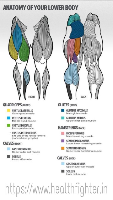
Quadriceps
1. The rectus femoris arises from the front of the pelvic bone.
2. The vastus medialis arises from the inner edge of the femur.
3. The vastus lateralis arises from the outer edge of the femur.
4. The vastus intermedius arises from the front surface of the femur and lies Underneath the rectus femoris.
The four heads merge, attach onto the patella (kneecap), and then insert via a single patellar tendon onto the tibia, just below the knee joint. The main function of the quadriceps is to extend the knee and straighten the leg. Because the rectus Femoris arises from the pelvic bone, contraction of this muscle also flexes the hip joint.
Hamstrings
The semimembranosus passes behind the inner aspect of the thigh, connecting to the upper tibia bone behind the knee. The semitendinosus passes behind the inner aspect of the thigh, connecting to the upper tibia bone adjacent to the semimembranosus.
All three hamstrings muscles span both the knee and hip joints. Therefore, they serve dual functions: flexion of the knee and extension of the hip.
Gluteal
Other muscles that move the hip joint include the following:
• Hip adductors (inner thigh): gracilis, adductor longus, adductor Magnus, and adductor brevis
• Hip abductors: tensor fasciae latae, gluteus medius, and gluteus minimus
• Hip flexors: Sartorius, iliopsoas, and rectus femoris
• Hip extensor: gluteus Maximus
Calves
The lower leg contains 10 muscles. The calf comprises two muscles. The gastrocnemius is the visible muscle of the calf. The 2 heads of the gastrocnemius medial and lateral arise from the rear of the femur bone, directly above the knee joint. The soleus arises from the rear aspect of the tibia and lies underneath the gastrocnemius.
The tendons of the gastrocnemius and soleus fuse to form the Achilles tendon, which passes behind the ankle joint and attaches to the calcaneus (heel bone). The calf muscles cause plantar flexion of the ankle, the movement required for standing on tiptoes.
The relative contribution of the two calf muscles depends on the angle of knee flexion. The gastrocnemius is the prime mover when the leg is straight, and the soleus becomes more active as the knee bends.
Note that the gastrocnemius crosses both the knee and ankle joints and therefore serves a double function: knee flexion and ankle flexion.
The following are other lower-leg muscles:
• Ankle extension (dorsiflexion): tibialis anterior
• Ankle eversion: peroneus longus and peroneus brevis
• Ankle inversion: tibialis posterior
• Toe flexors and extensors: flexor digitorum longus, flexor hallucis longus, extensor digitorum longus, and extensor hallucis longus
Lower Body
 Reviewed by HealthFighter
on
February 29, 2020
Rating:
Reviewed by HealthFighter
on
February 29, 2020
Rating:
Reviewed by HealthFighter
on
February 29, 2020
Rating:


No comments:
Please do not enter any spam links in the comment box